Overview
Imaging of biological specimens plays an essential role in research across many scientific disciplines. It enables measurements and visualization of complex biological systems with a high spatial and temporal resolution, to generate and further test scientific hypotheses.
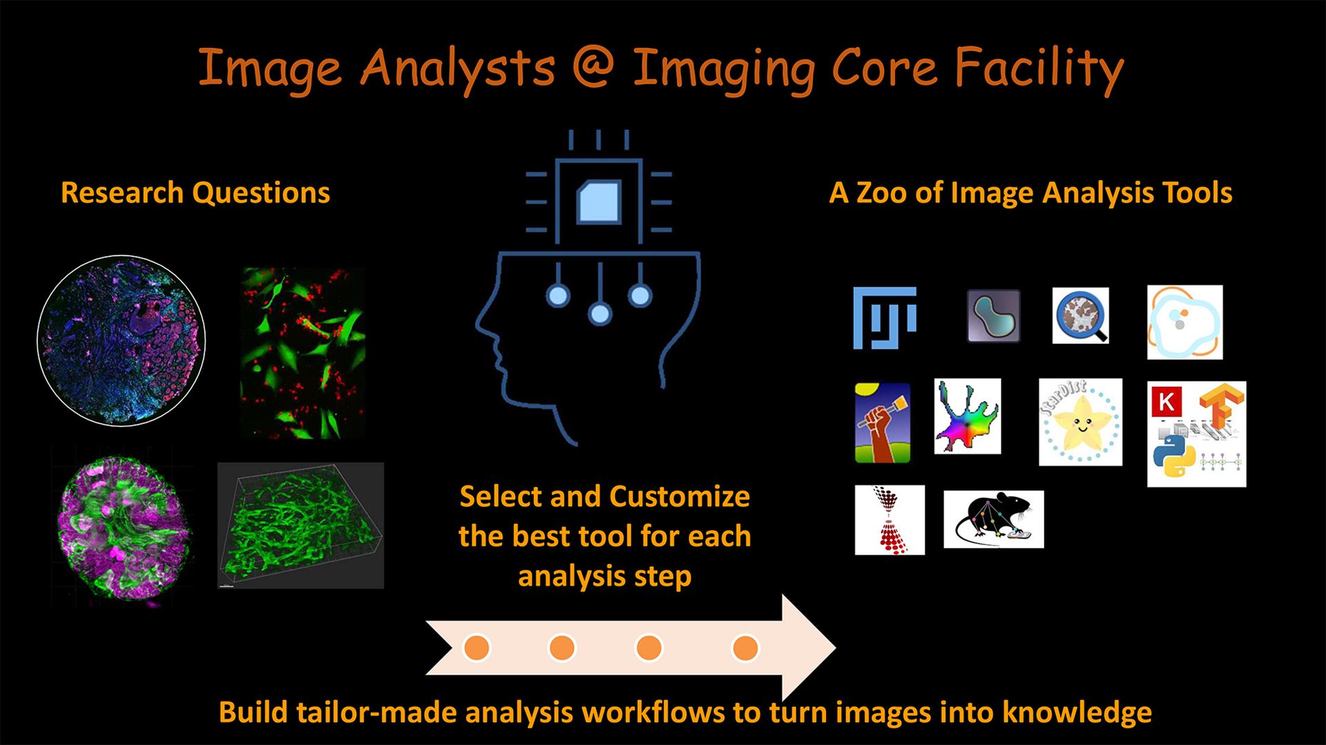
The task of bioimage analysis is to “enable [computers] to automatically distinguish between relevant and irrelevant image information and to recognize and model spatial or temporal patterns of interest that could confirm or refute the hypotheses underlying the given experiment through quantitative analysis.” [4]
To fulfill this, image analysis services in academic facilities address all issues related to post-acquisition of biological imaging data from various imaging modalities. This covers image visualization, image processing and quantitative image analysis.
Many image analysis software platforms and packages are available. An expert bioimage analyst will help choose and correctly utilize the right tool or combination of tools to build a workflow that will achieve the quantification goals. When needed, custom modules and tools will be developed.
We support researchers with:
- Quantification projects and workflow development
- Commercial software licenses
- Dedicated image analysis workstations
- Courses and individual training for general image analysis concepts and for commercial and open source image analysis software.
It is highly recommended to consult with a bioimage analyst at the beginning of a project to discuss experimental design related to image analysis.
Selected References
- "Hitchhiker’s Guide through the Bio-image Analysis Software Universe
- Image.sc
- Image analysis training Resources by NEUBIAS Academy @ Home
- Meijering, Erik, et al. Imagining the future of bioimage analysis. Nat Biotechnol 2016.
- Introduction to Bioimage Analysis