Overview
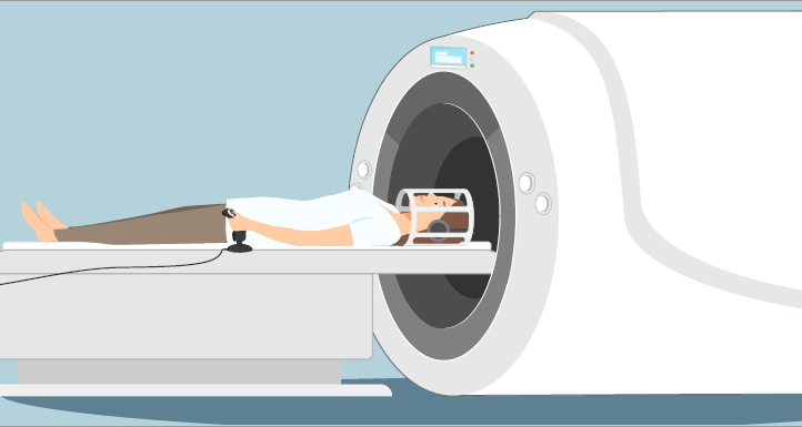
Magnetic resonance imaging (MRI) is a noninvasive imaging tool for pre-clinical and clinical research. It provides highly detailed images (sub millimeter spatial resolution) of the tissues in the body with many different contrast mechanisms. MRI allows mapping in 3-dimensions of anatomical and physiological features including: blood flow, perfusion, water diffusion, interstitial fluid pressure, vessel permeability, characterization of metabolic and biochemical processes by means of spectroscopy (MRS), as well as mapping of metabolic information (chemical shift imaging), and more.
Functional MRI (fMRI) is an MRI technique that allows measuring and mapping brain activity. The method is being used in a growing number of studies to map and learn about brain processes of sensory stimuli, language, memory, emotions, and social skills by challenging the subject with simple or complex stimuli or tasks while inside the magnet. The method also allows mapping and understanding the relationship between the structure and function of the various brain areas in healthy subjects and in various pathologies.

|
Selected Applications
|
Outcome
|
|
T1w and T2w Imaging
|
Anatomic structural MRI
|
|
BOLD, FMRI
|
Brain activity mapping
|
|
DWI/DTI
|
Water diffusion mapping and tissue architecture
|
|
Perfusion
|
Blood flow and blood volume mapping
|
|
MRS / CSI
|
Metabolic spectroscopy and imaging
|
|
SWI
|
Mapping of tissue susceptibility changes caused by various compounds, such as blood, iron and calcium
|